Scientific imaging in the fight against Covid-19
Coronaviruses are prolific across the world in the form of cold and flu germs, and have even caused global health issues in the shape of SARS (2003) and MERS (2012). However, nothing has been seen to date that matches the contagiousness and potential danger posed by Covid-19. Scientists are now sharing data in an unprecedented fashion to speed up how we understand and confront this novel threat. Imaging techniques are frontline weapons in the battle against the virus.
RNA imaging enables the development of test kits
Following the initial outbreak in China, the genetic makeup, or genome, of the virus was made globally available within days, compared to the months that it took in the case of SARS. Understanding the Ribonucleic Acid (RNA) molecule of the virus is paramount to knowing how it is transmitted and how it might be tackled. Fixed-cell imaging techniques are being widely replaced by live-cell technologies in RNA imaging. This involves the use of time-lapse microscopy which may be phase contrast, quantitative phase contrast or fluorescence microscopy, or holotomography. Phase contrast and fluorescent microscopy require cells to be stained in order to visualize them, which can of course be detrimental to the structure and survival of the cell.
One team based at the Simon Fraser University, Canada, is using their established RNA imaging technologies to develop Covid-19 testing kits. They are extending the scope of their Mango technology to detect the Covid-19 coronavirus, a single strand RNA virus. The Mango system uses an RNA sequence modified to contain an aptamer that binds to a fluorescent dye, which glows as it becomes excited. The RNA molecules carrying the aptamer are clearly visible under a fluorescence microscope, allowing rapid detection of virus particles.
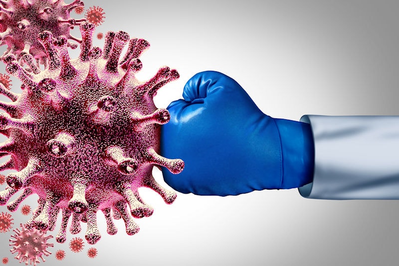
Attacking the virus
Experts in CRISPR sequencing at the Donnelly Centre for Cellular and Biomolecular Research, also in Canada, have been looking at how to remove gene fragments from cells which could result in an effective treatment or even vaccines for Covid-19. Their latest technique involves using a specific enzyme (Cas13) to recognize, track and cleave, or “knock down”, particular RNA molecules in human genomes and may be put to use in destroying the Covid-19 virus. Previous research with the enzyme Cas9 has been shown to eliminate HIV in living animals, slow the growth of cancer cells and even change the color of a flower. A high-speed atomic force microscope (AFM) is used to image the process as this scanning probe microscope is able to visualize dynamic biomolecular processes at sub-second resolution.
Tracking the spread with next-generation sequencing
A consortium of researchers in the UK are being given access to viral nucleic acid from patient samples who have tested positive for Covid-19. Led by the Wellcome Sanger Institute, one of the world’s most advanced centers for genome sequencing, scientists will use next-generation sequencing (NGS) to analyze the samples using advanced imaging and algorithms. This high-throughput, massively parallel method will be used to provide insights into the geographical spread and evolution of the virus. Such methods have already informed us that the spread of Covid-19 to Iran originated from just one person, but it was likely to have entered the UK from four sources.
Keeping us safe with thermal imaging
Early detection of symptoms is key in containing the spread of Covid-19. Thermal imaging has been employed by border staff around the world and even in public areas in China to identify potential carriers of the disease. Baidu have developed an AI system which is monitoring travelers through the Qinghe Railway Station in Beijing. Computer vision cameras with infrared sensors can capture and process images of up to 200 people every minute and flag those who have a temperature of over 37.3°C. US thermal imaging manufacturer FLIR have reported “strong demand” for their thermal cameras which are used in Elevated Body Temperature Screening. DJI, one of the world’s largest drone manufacturers, has found that adding a cotton swab to their drones carrying a thermal camera decreased the margin of error from 4-5°C to just 0.5°C by acting as a calibration reference for the camera.
The Covid-19 pandemic has left the world reeling but we’re proud to be part of an industry which is leading the fight against the virus and supporting the science behind its control. View our computer vision products online and contact us to see how our products are advancing scientific research globally.