Medical Imaging Techniques – How do they work?
A wide range of imaging techniques are used to enhance medical diagnoses and treatments. Some of these are well-established and have been supporting the sector for years, but there are always new and innovative methods being developed. Here, we look at some of the most commonly used methods.
Ionizing Radiation Imaging
Ionizing radiation–based medical imaging refers to techniques that use the high-energy x-ray radiation to visualize the inside of the human body.
- X-ray Imaging (radiography) is perhaps the most universally known radiation imaging technique. It is a projection-based method that uses high-energy electromagnetic radiation that is sent through parts of the body to capture the internal structure. A detector produces an X-ray image, which is a 2D intensity map where pixel brightness corresponds to the attenuation of X-ray photons as they pass through materials of varying density and atomic composition. Dense tissues like bone absorb more radiation and appear bright, while soft tissues and air-filled spaces appear darker. Modern approaches leverage deep learning for automated diagnosis, bone-fracture detection, and disease screening.
- Computed Tomography (CT) is a variation of X-ray–based imaging that produces cross-sectional images (“slices”) of the body. An X-ray source rotates quite fast around the patient while detectors measure the reduction in signal as X-rays pass through tissue, known as attenuation. The system collects hundreds to thousands of projections from different angles. These are reconstructed into a 3D representation of the patient using algorithms (for example, filtered back projection or iterative reconstruction). CT systems require high data throughput, precise timing, and efficient memory handling to process large volumes of projection data in real time.
- Fluoroscopy is a real-time X-ray imaging technique that produces continuous image sequences or video of internal structures. It is used to observe swallowing, catheter insertion, joint movement, cardiac motion, contrast flow etc. An X-ray source emits a steady or pulsed beam, and a detector captures transmitted radiation to form frames at high rates. Image intensifiers or flat-panel detectors convert X-rays into visible signals. Systems must handle low-latency acquisition and processing to support live guidance during procedures. Dose management is critical, so pulsed imaging and temporal filtering (combining multiple frames to reduce noise) are commonly used to balance image quality and radiation exposure.
Light-based Imaging Techniques
Light-based imaging methods use optical radiation (typically visible to near‑infrared light) and mechanisms such as reflection, scattering, fluorescence, and interference to visualize tissues from cellular to macroscopic scales, often with high spatial resolution.
- Optical Coherence Tomography (OCT) uses light waves to create high-resolution cross-sectional images of biological tissues and is used commonly in ophthalmology for imaging the retina. OCT works by measuring the echo time delay and intensity of backscattered light, allowing for precise visualization of tissue layers and structures.
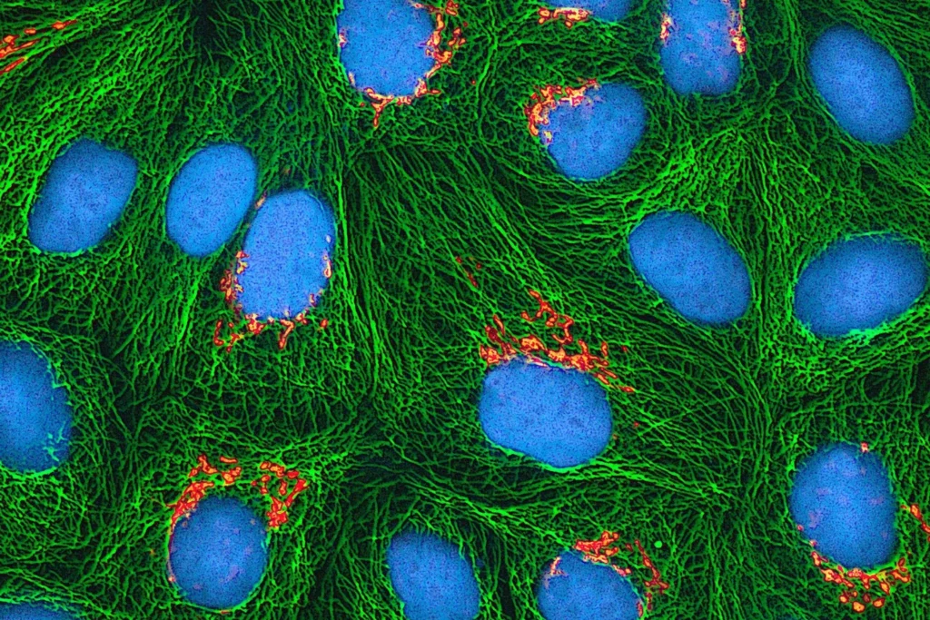
- Confocal Microscopy uses focused laser light and a pinhole (an optical aperture that blocks out-of-focus light) to create high-resolution images of thin tissue sections or cells. By scanning point-by-point and rejecting scattered light, it produces sharp images with good depth discrimination. Confocal microscopy is widely used in research and increasingly in clinical applications such as dermatology and ophthalmology. It is different from OCT because it provides very high lateral resolution but typically requires closer, more localized imaging rather than deep tissue penetration.
- Fluorescence Imaging is a light-based technique that uses fluorescent dyes or markers to visualize biological structures and processes. A light source (often a laser) excites these molecules, causing them to emit light at a longer wavelength. The emitted signal is captured by detectors to form images. This method provides high sensitivity and can highlight specific cells, proteins, or biomarkers when paired with targeted contrast agents. It is widely used in research, molecular imaging, and image-guided surgery.
Other Common Medical Imaging Methods
Although ultrasound cannot produce high-quality images in air-filled or bone-obstructed regions, its cost-effectiveness, portability and safety make it a widely used diagnostic tool.
- Ultrasound imaging uses high-frequency sound waves to create real-time images of internal body structures. Here, instead of electromagnetic radiation, acoustic pulses are transmitted into tissue. The echoes that bounce back from boundaries between different tissue types are then detected. The timing and strength of these echoes are processed into 2D or 3D grayscale images, similar to how depth sensors reconstruct 3D scenes. Ultrasound’s main advantages are real-time imaging speeds, safety, and the ability to measure blood flow using Doppler techniques.
Magnetic Resonance Imaging offers excellent soft‑tissue contrast, multiplanar 3D imaging, and no ionizing radiation, making it ideal for brain, spine and joint assessment.
- MRI uses a strong magnetic field and radio waves to generate detailed images of the internal structures of the body. However, it is expensive, slow, sensitive to motion, and unsuitable for some patients with metal implants or claustrophobia. Functional MRI (fMRI) measures and maps brain activity by detecting changes in blood flow and oxygenation, providing an indirect marker of neural activity.
Compact Imaging Solutions
Tools designed around miniaturization and portability can still offer significant diagnostic capability. Techniques such as Endoscopy use flexible probes with integrated light and cameras to access internal organs with minimal invasiveness. It is widely used in gastrointestinal and respiratory applications.
Capsule endoscopes are swallowable imaging devices which provide even less invasive visualization of the gastrointestinal tract. They transmit images wirelessly as they pass through the body.
Advances in wearable imaging technology enable continuous or point-of-care monitoring, integrating lightweight sensors and low-power imaging modules for applications such as wound assessment or vascular monitoring. For example, Singapore-MIT Alliance for Research and Technology (SMART) has launched its Wearable Imaging for Transforming Elderly Care (WITEC). This tiny ultrasound system conducts cardiovascular imaging for continuous and real-time monitoring and diagnosis of chronic conditions such as hypertension and heart failure.
These systems emphasize low power consumption, efficient data transmission, and compact sensor integration. Compact imaging is expanding access to diagnostics while shifting imaging closer to the patient.
An Arsenal of Imaging Tools
Medical imaging techniques, leveraging radiation, light, sound, and more, enable non-invasive visualization of the human body. Although there may be varying trade-offs in resolution, depth, safety, and speed, today’s array of imaging tools support accurate diagnoses, treatment, and research across modern healthcare fields.
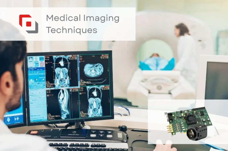
Our high-speed, high-resolution frame grabbers and embedded systems support several medical systems and are currently used in applications such as ophthalmology and oncology. Take a look at how our products support the medical sector.