Bringing CCD performance to CMOS cameras
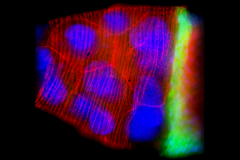
Active Silicon partners closely with a number of camera manufacturers and specialists in scientific imaging for medical and life science applications. Our range of FireBird Camera Link frame grabbers offers advanced functionality and reliable operation, allowing camera manufacturers to reach new limits in, amongst other things, microscopy. Epifluorescence microscopy provides a particular challenge to imaging due to low light levels, high signal-to-noise levels and because the emitted light often fades during the microscopy process. In fluorescence microscopy, a specimen is illuminated using light of a specific wavelength, triggering fluorescence of certain components. Typically, biological specimens don’t fluoresce themselves, rather specific structures in the specimen are dyed with chemicals, called fluorochromes, that fluoresce when excited by the light, thereby making it possible to identify very specific cellular components and impurities to a greater degree than with other microscopic methods. It is possible to dye different structures with different color emitting dyes, as can be seen in the accompanying image.
The picture shows the egg chamber of a Drosophila. The fruit fly lends itself particularly well to studying cell function and development in biomedical research, in this particular image the development of eggs (oogenesis) was studied. The DNA in the individual cells is shown in blue, the F-actin cytoskeleton in red and the mRNA at the transition to the main body area in green. The fluorochrome dyes used were DAPI, Rhodamine, and GFP.
A major advantage of modern CMOS cameras for microscopy is the ability to operate in low light environments, which means the cells are less likely to suffer deterioration from photobleaching. The combination of high quantum efficiency and low noise is allowing advances in imaging beyond the capabilities of CCD cameras, and enabling improved research on cell development over time.
One of our customers has been able to develop a digital CMOS camera with twice the speed, three times the field of view and drastically less noise than the market-leading CCD cameras. The Camera Link CMOS camera, once connected to our Camera Link frame grabber, allows images of 4 megapixels and 16-bit to be transferred to a host PC at 100 frames/sec. The real-time processing and high data-rate performance are matching the more expensive CCD units and offering a more affordable solution for bright-field, fluorescence and a range of other microscopy techniques.
Other applications of this camera include super-resolution microscopy, TIRF microscopy, ratio imaging, FRET, High-speed Ca2+ imaging, Real-time confocal microscopy and light sheet microscopy.
Active Silicon is proud to support our customers developing these, and other, revolutionary imaging processes.