CoaXPress frame grabbers support time-lapse imaging for life science
Time-lapse imaging is the practice of taking thousands of images of a subject over a course of time to monitor changes to that subject. It is widely used in life science to monitor growth, movement and development of cells. Film frames are captured at a lower frequency than they are viewed, to allow scientists to view long-term changes in a short period.
Steps to successful time-lapse imaging
Planning: It’s important to carefully plan and prepare time-lapse imaging experiments to be able to get evaluable results. For example, the best possible conditions to keep the living cells alive and active under the microscope need to be established and maintained. The microscopic set-up is crucial, different microscopes and techniques will be suitable to answer different scientific questions. Also of great importance is to determine the sampling rate – the rate at which images should be acquired. This should be often enough not to miss crucial steps, but the light sensitivity of living cells will limit the number of possible samplings.
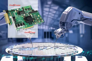
Image acquisition: Camera selection will be made based on factors including sensor resolution, frame rates and, for time-lapse in particular, high quantum efficiency and extremely low readout noise as these experiments usually run in low light conditions and depict structures with low contrasts. Using a CoaXPress camera, such as Hamamatsu’s scientific qCMOS 4xCXP ORCA-Quest camera, provides exceptionally high sensitivity and enables extremely fast image transfer of high resolution images to a CoaXPress frame grabber – the latest acquisition cards enable a maximum data rate of 50 Gbps to be achieved – combined with precise triggering capability.
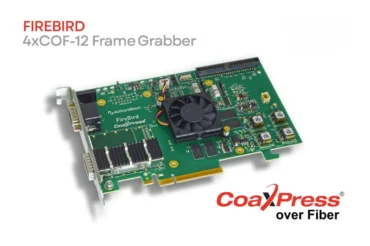
Analysis of images and data: Various software applications are available to support advanced data analysis, such as NI’s LabVIEW or MathWorks’ MATLAB. Both of these, and several others, are compatible with Active Silicon’s FireBird CoaXPress frame grabbers. They enable the rapid collection and analysis of data, leading to quickly-built test environments and easy-to-replicate programs.
Drawing conclusions: Being able to design, test and confirm theories relies heavily on accurate, credible and repeatable data. Time-lapse imaging is an application which, by its very nature, produces different results each time as well as huge amounts of image data, so automated object tracking and algorithms are used to look for patterns in data from which conclusions can be successfully drawn. Therefore, a wide library of data may be required.
Applications for time-lapse imaging
Researching cell division
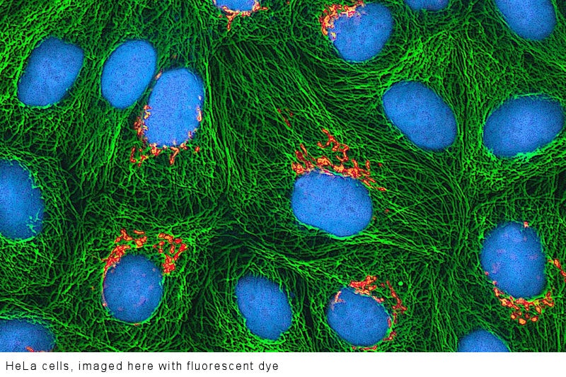
Time-lapse microscopy is used to view the dividing of HeLa cells – cells that carry mutations in their DNA which cause them to continually divide rather than ever die. Removed during a biopsy from Henrietta Lacks, from where their name derives, these cancerous cells are known as immortal and provide a crucial example of an immortalized cell line. In fact, the first HeLa cell to be discovered in 1951 has daughter cells still in existence today which are sold for scientific research. Credited for advancements in vaccines and genome mapping, HeLa cells have played an important role in scientific research. Several Nobel prize winning research projects have depended on the use of this oldest and still one of very few immortal human cell lines.
Monitoring cancer treatment
Recent developments have allowed scientists to stain cells without destroying them and these are viewed using a phase-contrast microscope in a process known as live cell imaging. Cell tracking is used to determine the reaction of cells to different cancer treatment medications. In such instances, time-lapse imaging is generally carried out over a 96 hour period, although several weeks are possible. Cell growth, cell cycle progression, damage and cell death can be monitored. In a similar way, monitoring over a longer period allows oncologists to understand how cancers grow, providing invaluable data for novel research.
Boosting IVF success
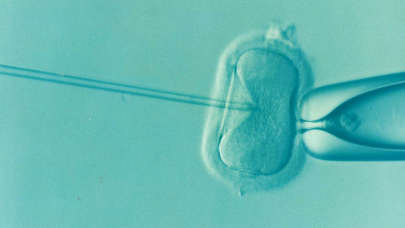
Time-lapse incubation and imaging is used in IVF procedures to help identify the embryos most likely to successfully develop into fetuses. Usually, embryos have to be removed from the incubator to be examined under a microscope; time-lapse imaging now allows doctors to leave the embryo in situ in the incubator and monitored over time to determine the sample with the optimum characteristics and cell development for implantation. Newly designed “EmbryoScope” incubators encompass a built-in camera unit connected to a microscope system and advanced software removes the guesswork from selecting suitable embryos.
Quality components deliver quality data
While time-lapse imaging may have traditionally not required high data rates or frame rates, modern high-resolution sensors and fast exposures settings still require very high speed readout rates and these are getting faster all the time. Fast action life science analysis may also require high frames rates even while it’s still considered as time-lapse imaging.
Active Silicon CoaXPress frame grabbers in combination with powerful scientific cameras provide imaging solutions which are reaching new horizons in life science research. Contact our team to see how to bring your microscope systems to the forefront of scientific exploration.