Developments in Medical Imaging

Complex computer vision products and software have been used in medical imaging for decades but the scale at which these are developing is unprecedented.
We take a look at some examples of how imaging for medical applications is enabling superior healthcare across the globe.
Adapting traditional imaging techniques for novel applications
Radioactive gold for monitoring cancer treatment
In Japan, researchers from Waseda University have been working on a technique which uses neutron activation to transform gold particles into a radioisotope of the precious metal. Gold nanoparticles (AuNPs) have been found to be a promising carrier for targeting drug delivery, especially in the treatment of some cancers.
This new imaging method enables better long-term tracking of the nanoparticles in the human body. For activation of the AuNPs, the researchers irradiated regular gold nanoparticles (197Au) with neutrons, converting the stable form into a radioactive form (198Au). The radioactive 198Au emits gamma rays, which are detectable from outside the body.
Detecting “hot” tumor cells with phasor thermography
Another research team based at the Georgia Institute of Technology has modified a thermal imaging platform to assess a patient’s vital signs for early detection of disease or other ill health conditions.
To avoid the “ghosting effect”, where broad and overlapping emission spectra can make details imperceptible, the team have employed phasor analysis – an established numerical method for reducing fluorescence lifetime data complexity. The phasor thermography (PTG) device works by taking ten separate infrared images, each capturing a slightly different part of the long-wavelength infrared spectrum. Using a mathematical technique called thermal phasor analysis (adapted from signal processing) it can combine these images to reveal tiny surface details less than a millimeter in size.
This level of precision makes it possible to see subtle temperature differences, clearly distinguishing between facial skin, thick scalp hair, and even the finer hair of the eyebrows. This type of thermography can assist in early detection of abnormal cell activity associated with early cancer. Such cells use more oxygen to develop and reproduce and thereby expel increased heat.
Industrial techniques tailored for medical imaging
Line scan cameras to examine skin
Line scan imaging, widely used throughout industrial inspection, hasn’t generally been associated with medical imaging – until now. Line scan imaging uses a single-row array of sensors to scan the subject one line at a time, recording visual data sequentially along a narrow path. The collected lines are then stitched together to create a complete image.
Researchers at Brno University of Technology in the Czech Republic have developed a line scan device specifically for imaging the fingers, hand and even arm. This IMVE article explains the device in detail, but results should be able to examine skin conditions such as eczema and monitor anomalies and growths.
SWIR fluorescence improves medical image quality
Another technique used increasingly in industrial inspection is Short-Wave Infrared (SWIR) imaging. This method allows inspection of materials that interact differently with SWIR light compared to visible light, allowing for the identification of specific materials or the analysis of their composition.
Scientists at the UCL Great Ormond Street Institute of Child Health have trialed using SWIR fluorescence to improve the quality of images to distinguish between cancerous tumors and healthy tissue during preclinical tests. SWIR has been found to enhance the quality of molecular images in real time and may in the future be used during procedures to help surgeons remove cancerous tissue more precisely.
The inevitable rise of AI to progress medical imaging
The medical sector is among one of many benefitting from massive global investment in AI. With healthcare professionals under increasing pressure, there are clear advantages to applying advanced machine learning and AI techniques to collating and analyzing medical data.
AI determines the microsatellite status of colon cancer
In the crusade against colon cancer, AI is being blended with infrared imaging to help create more effective therapies. In order to tailor therapies to individuals to deliver the very best outcomes, very accurate diagnoses are required. A team from the Centre for Protein Diagnostics (PRODI) at Ruhr University Bochum have used infrared imaging to measure the genomic and proteomic composition of tissue to provide information at the molecular level and applied AI to determine the microsatellite status, that is, to identify whether a patient will respond positively to immunotherapy. This process has been developed to give results in about an hour, saving a huge amount of time compared to traditional immunostaining methods, and enabling treatment to begin sooner.
Dracula neural network to improve radiation therapy
Dracula is the name given to a Deep Radial Convolutional Neural Network developed by The Institute of Cancer Research and The Royal Marsden NHS Foundation Trust to improve and accelerate the delivery of radiation therapy. The algorithm reconstructs MRI images of moving tumors, allowing radiation treatment to be targeted at cancerous tissue while avoiding healthy organs, even during the respiratory phase (i.e. when the patient’s breathing causes movement).
The required reconstruction of 4D MRI images takes less than a minute with Dracula, compared to the previous methods which took several hours. The results mean that higher doses of radiation can safely be delivered directly to the tumor with lower risk of damaging surrounding tissue.
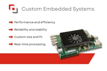
Experts in designing computer vision products for medical applications
Active Silicon has been producing components for medical imaging devices for over 30 years. Our embedded systems, frame grabbers and autofocus-zoom cameras are all used in medical imaging applications, from ophthalmology to cancer treatment. We understand the importance of high-quality images delivered in real-time in products that will operate reliably for many years.
Take a look at what we’ve done in the past for the medical sector, and contact us to see how we can assist your imaging application.