The role of computer imaging in post-operative practices
We’ve looked at the use of imaging in pre-operative and real-time surgical situations. Medical imaging also plays a vital role in collecting data for post-operative analysis.
Implant surgery is one area where there’s an obvious use for imaging after the procedure, to make sure plates and devices have been correctly positioned, but the case for post-operative imaging is a strong one in many other fields.
Imaging in pathology
Another familiar use case for post-operative medical imaging is pathology. This, put simply, is the study of disease, often enhanced through the analysis of surgically removed organs and tissue. Samples are then examined using a combination of techniques including microscopy and digital pathology. Digital pathology uses scanning procedures which convert glass slides to high-resolution digital images which can be viewed and interpreted more accurately than under a microscope alone. As well as providing clear and adaptable images, the resulting pictures can be shared widely and lend themselves to advanced processing and AI techniques.
Inify Laboratories provides image enhancement for 2D and 3D medical devices. They combine state-of-the-art software with AI to support game-changing ultrasound, Magnetic Resonance Imaging (MRI), radiography and mammography. Their INIFY® Prostate Screening is an AI-powered tool that provides pathologists with valuable decision-making support when identifying cancer in prostate biopsies. It claims to produce faster and more accurate results for pathology than traditional microscopy. The tool uses a high-resolution multiplex immunofluorescence overlay to build a reliable and precise algorithm.
Identifying post-surgical complications
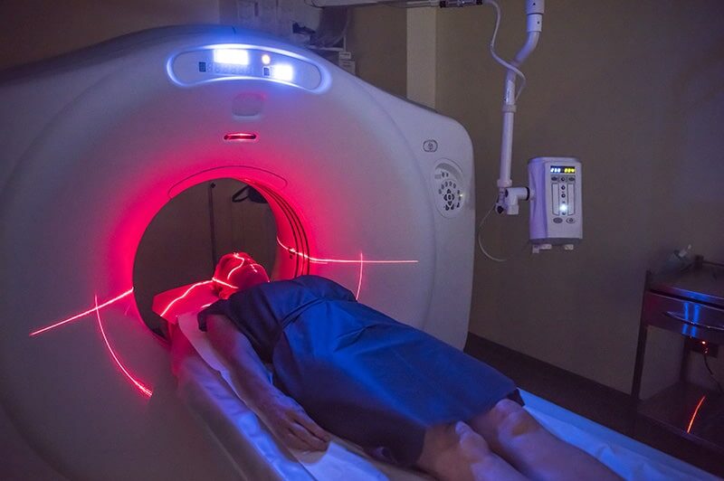
Chronic subdural hematoma (CSH) is bleeding between the skull and the surface of the brain caused by a head injury or trauma and may present as headaches, confusion, difficulty in speaking and loss of balance. The size of the hematoma, or bleed, may increase over a number of weeks in some cases and can often reoccur. Treatment generally consists of drilling a small burr hole in the skull, or trepanning, to allow blood to escape and relieve pressure on the brain. Computed tomography (CT) scans are essential for monitoring a patient after the procedure, allowing surgeons to check for further build-up of blood. If a hematoma builds up again, rapid detection and treatment can make the difference between life and death. Cranial imaging is an essential component of patient recovery following brain surgery.
Vision in Physiotherapy
Ultrasound is commonly used in post-trauma recovery plans due to its relatively low cost and wide availability. Ultrasound imaging uses reflected sound waves to produce 2D images of soft tissue in a non-invasive and low-risk procedure. This method of scanning is suitable for imaging muscles, fluids and tendons in superficial injuries. Where muscles and tendons have been cut or torn during injury or surgery, regular ultrasound scans can be used to monitor the repair of tissue and support a patient’s physiotherapy rehabilitation plan, particularly following hip or knee replacement surgery.
Companies such as Clarius are making vision available to a huge swathe of physiotherapists through their handheld ultrasound devices.
MRI involves measuring the response to a pulsed radiofrequency current in a strong magnetic field and can also play a part in post-operative physiotherapy. This technique produces higher-resolution images of deeper organs and tissue and is more widely associated with imaging nerves, the spinal cord and brain. The associated large, specialist equipment and the need for patients to remain completely still make MRI imaging more suited to clinical rehabilitation than to physio-on-the-go situations.
AI is making an appearance in this field too, and the Chartered Society of Physiotherapists (CSP) have written an informative blog covering the advantages and limitations of AI in physiotherapy.
Vision for every medical application
Computer vision and medical imaging are cornerstones for many surgical procedures, but their use doesn’t stop after the operating theater lights are switched off. Imaging is paramount for managing post-surgical complications and rehabilitation plans.
We have a range of computer vision products and embedded systems which are certified for use in surgical settings and medical procedures. Speak to our experts to see which one will deliver world-leading imaging to your particular medical application.