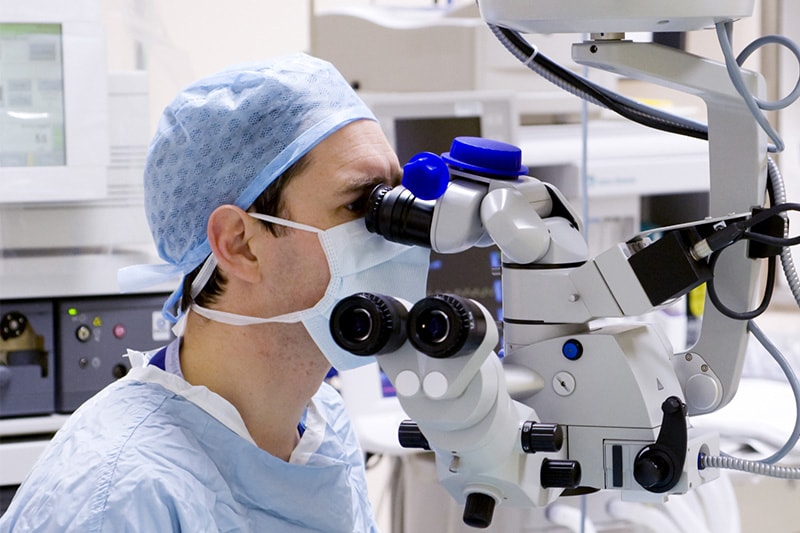
Imaging surgical procedures at the critical moment
Last month, the global market for AI-based clinical applications for use in medical imaging was forecast to reach nearly US$1.5bn by 2024[1]. We looked at pre-operative medical imaging in September (you can read our blog here.). There are also fascinating developments being made in the imaging of real-time surgical procedures, and we’ve gathered several innovative case studies below.
Medical imaging at your fingertips when it matters most
A project named “Data Acquisition, Analysis, and Visualization for 3D Computer Vision Surgical Robotics” is being developed at the Centre for Innovation in Information Visualization and Data Driven Design (CIVDDD) in Canada. It is set to advance the use of computer vision and data visualization for image-guided robotic surgery. A 3D visualization of the surgical procedure automatically relates images from pre-surgical plans to make a real-time comparison with how the operation is going according to the plan. This is then able to accommodate unexpected deviations should problems occur, and provides greater guidance to the surgeon during the procedure in the event of unforeseen events.
Where did I put that sponge?
Leaving a surgical sponge behind while conducting surgery is never a good thing, and more common than you might think – 1 in every 5,500 surgeries involves a retained sponge. Gauss’ Triton software uses an iPhone camera and AI to track and count every surgical sponge used, even when different types, sizes and standard of sponge are present. Gauss is even developing a machine learning solution to monitor the amount of blood loss during surgery. For that, images of used sponges and canisters are captured, and an estimate of blood loss is calculated in real time. The company claims that trials led to markedly less additional blood product being needed during cesarean sections, and a reduced recovery time in hospital.
Multispectral imaging plays its part
Quest Medical Imaging offer their Quest Spectrum®, a multispectral camera with processing equipment to visualize cancer cells and tissue differences that are not visible to the human eye. For example, tumours can be examined during surgery by injecting tumour-specific markers labelled with Indocyanine Green (ICG) into the patient some days before. When the markers have bound to the tumour, it becomes visible with fluorescence imaging. During an operation, the color image of the organ is overlaid with the image of the fluorescence signal to identify the tumour among the healthy tissue.
Active Silicon supports surgical success
Computer vision has long played an important part in advancing surgical procedures. Now, the combination of improved imaging, faster processing and pioneering AI is enabling visualizations in the operating theatre that reduce errors and improve success rates. We’re proud to be supplying leading-edge technology which is helping medical imaging evolve and transform the future of ground-breaking surgery. View our products online and get in touch to see how we can customize our hardware and software to suit the requirements of global medical imaging providers.
[1] https://www.prnewswire.com/news-releases/ai-in-medical-imaging-to-reach-1-5-billion-by-2024–301135370.html