Overcoming Image Processing Challenges in OCT Medical Imaging Systems
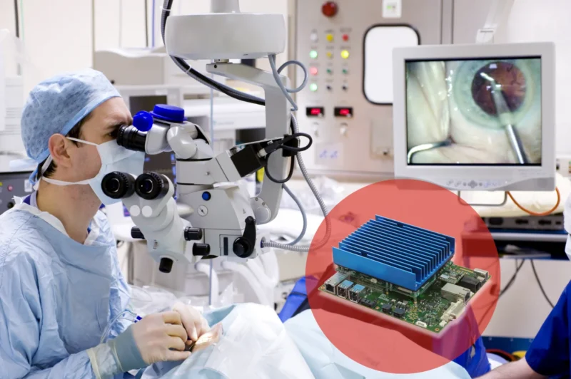
Medical imaging technology continues to advance rapidly, helping clinicians diagnose conditions earlier and with greater confidence. One technique that is seeing significant growth currently is Optical Coherence Tomography (OCT).
Widely used in ophthalmology, cardiology, dermatology, and research applications, OCT provides high-resolution cross-sectional images of biological tissue in real time.
As OCT systems become more sophisticated, equipment manufacturers face a growing challenge: how to process and manage increasing volumes of imaging data while maintaining image quality, system reliability, and compact device designs.
Different OCT Techniques
While there are several variations of OCT approaches, modern OCT systems are primarily based on spectral-domain and swept-source technologies. These have replaced earlier time-domain approaches in many clinical applications due to their higher imaging speeds and improved sensitivity.
Spectral-domain OCT (SD-OCT)
Spectral-domain OCT uses a broadband light source and a spectrometer to measure the different wavelengths of reflected light simultaneously. The basic process is:
- A beam of light is split between a reference path and the tissue being imaged.
- Reflected light from the tissue returns and combines with the reference signal.
- A spectrometer captures the interference pattern across many wavelengths.
- A mathematical process called a Fourier transform converts this data into a depth-resolved image.
Advantages of SD-OCT:
- Very high image resolution
- Excellent sensitivity
- Fast scanning – 20,000 to 40,000 scans per second
- Widely used in ophthalmology, particularly retinal imaging
Swept-source OCT (SS-OCT)
Swept-source OCT uses a different approach. Instead of capturing a wide range of wavelengths at once, it uses a tunable laser that rapidly sweeps through different wavelengths. During each sweep:
- The laser emits a changing wavelength of light.
- Reflections from the tissue are captured by a high-speed photodetector.
- The collected data is processed to create a depth profile.
- Multiple depth profiles are combined to form an image or 3D volume.
Advantages of SS-OCT:
- Much higher imaging speeds (100,000 to 400,000 scans per second)
- Greater imaging depth
- Better penetration through certain tissues
- Reduced sensitivity roll-off at greater depths
These characteristics make SS-OCT particularly attractive for applications such as deeper retinal imaging, cardiovascular imaging, and other areas where rapid acquisition of large volumes is important.
The Challenge: High-Speed Data Acquisition and Real-Time Processing
Modern OCT systems generate vast amounts of data. SD-OCT and SS-OCT technologies can capture hundreds of thousands of scans per second, producing detailed images that help clinicians identify subtle structural changes in tissue.
Plus, the challenge is not simply “more data”; it is that modern OCT systems are expected to capture, reconstruct, enhance, analyze, and display increasingly complex images in real time.
However, performance comes at a cost. Medical device designers must handle high-bandwidth sensor data, perform complex image reconstruction algorithms, and deliver real-time visualization without introducing latency. At the same time, systems must meet strict requirements for reliability, power efficiency, and regulatory compliance.
The challenge becomes even greater when integrating multiple imaging modalities, incorporating AI-assisted analysis, or developing portable and point-of-care diagnostic devices. Traditional off-the-shelf processing solutions can struggle to provide the flexibility and performance required for these demanding applications.
The Solution: Custom Embedded Vision Systems for OCT
Custom-designed embedded vision systems offer a powerful way to overcome these challenges.
By combining advanced image acquisition hardware with high-performance processing technologies, embedded vision platforms can be tailored specifically to the requirements of an OCT application. This allows medical device manufacturers to optimize every stage of the imaging pipeline, from sensor capture through to image reconstruction and display.
FPGA-based processing can be used to manage high-speed data streams and perform deterministic real-time operations. GPUs and specialized AI accelerators can support advanced image enhancement, segmentation, and analysis algorithms. Together, these technologies enable rapid processing of large OCT datasets while maintaining exceptional image quality.
A custom architecture also allows designers to balance performance, power consumption, physical size, and cost. This is particularly valuable for portable imaging systems where space and thermal constraints are critical design considerations.
Enabling Next-Generation OCT Innovation
The flexibility of custom embedded vision systems extends beyond performance improvements. Manufacturers can integrate precisely the interfaces, sensors and processing capabilities required for their application, reducing unnecessary complexity and simplifying long-term product development.
As OCT technology evolves, system architectures can be designed to accommodate future enhancements, including higher-resolution sensors, faster acquisition rates and machine learning-based diagnostic tools. This scalability helps protect development investments while supporting innovation throughout the product lifecycle.
Building Medical Imaging Systems for the Future
Custom-designed embedded vision systems provide a practical and scalable solution. By delivering optimized image acquisition, real-time processing, and flexible system architectures, they help medical device manufacturers create the next generation of platforms capable of meeting both clinical and commercial demands.
Talk to us about our custom design skills and embedded vision solutions. We’re already supplying to the ophthalmological sector and can assist with image acquisition across many medical fields.