High-resolution digital slide scanners support improved pathology results
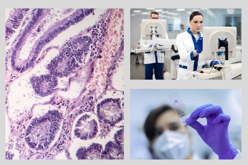
Pathology is the study of diseases and how they affect the body, although in modern healthcare the term is more commonly used to describe the field of medicine concerned with diagnosing disease. A variety of tools are available for pathologists to help collect and analyze data, as well as share research and developments. Digital slide scanners are one device in a pathologist’s armory that is used to capture very high-resolution images of tissue samples, such as those taken during a biopsy or surgical procedure. We’ve previously published a case study on using frame grabbers in digital scanners for faster breast cancer diagnosis.
Creating digital slides
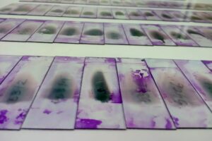
Tissue samples collected in a biopsy or other procedure have been used for a while to successfully diagnose and monitor diseases and medical conditions. Careful handling and optimal preparation of the sample is paramount to obtain high quality samples, this is even more critical when using automated systems like slide scanners. The preparation typically involves embedding the sample in paraffin wax, which helps to preserve the tissue and make it easier to slice into thin sections, which is then done using a microtome. The sections are typically between 2-5 microns thick and are mounted onto glass slides.
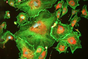
These tissue sections are then stained using special dyes which help to highlight different structures within the tissue. For example, Hematoxylin, which stains proteins blue, and Eosin, which stains proteins pink, are frequently used alone or in combination to create more defined contrasts. In fluorescence microscopy, various fluorescing dyes are available to highlight different types of cell structures. The staining process can take several hours to complete.
Once the tissue sections are stained, they are ready to be imaged. Digital slide scanners, like the NanoZoomer series from Hamamatsu, the Aperio range from Leica or the PANNORAMIC Diagnostic Scanners from 3DHISTECH are used to capture very high-resolution images of each tissue section. The scanner moves across the slide, capturing images of each field of view. The resulting images are saved as digital files. Specialized software, usually supplied by the scanner manufacturer, presents the files to the operator or surgeon to enable simple examination and manipulation of the images.
Before the digital slide is considered complete, it must undergo quality checks to ensure that the images are of sufficient quality for diagnosis. This can involve checking for issues that could affect the accuracy of the diagnosis, for example, debris, bubbles under the coverslip or artifacts on the tissue sample may cause the slide scanner to focus on these instead of the sample itself, and vital areas of the sample may then be out of focus. The digital slide is then stored in an image database where it can be accessed by pathologists and other healthcare professionals.
Analyzing pathology image data
Specialist software, such as MATLAB from Mathworks, can be used to manage medical image databases and enables segmentation, classification, registration, and 3D reconstruction of image data.
Data is analyzed to identify trends and patterns, or using AI to develop predictive models based on the data. Advances in the use of AI in medicine is helping healthcare professionals to identify potential diagnoses, monitor patient outcomes, and improve treatment protocols. AI for pathology requires large datasets for training and these can be created using whole slide images (WSI). The vast number of WSIs required for machine learning is now more readily available due to the increased number of digital slides produced every day.
Pathology data may need to be shared with other professionals, such as primary care physicians or specialists. This can help to ensure that patients receive coordinated care and that all healthcare providers have access to the same information about a patient’s health. If image data is digitized, it can be accessed by multiple parties regardless of where they’re based, meaning patients can have access to specialist care from anywhere in the world.
Digital slide scanners for digital pathology
Digital scanners can capture images at extremely high resolutions, allowing pathologists to see details that might be missed with other forms of microscopy. High-quality digital images of tissue samples are vital for accurate examination of small, delicate or complex samples, such as those from the brain or other organs.
Digital storage makes for easier re-access too – pathologists can view samples at their desk rather than having to retrieve a slide from storage and re-examine it under a microscope. Additionally, biological samples will inevitably deteriorate over time while a digital slide records the structure, colors and other features right after sampling and preserves this quality.
Imaging digitally also enables fast throughput, and thousands of slides can be loaded on several trays at one time and scanned by high-speed scanners. Such scanners are often optimized for imaging a particular sample type and imaging modes can be pre-set to acquire the best quality image for this type of tissue. With barcode information being read and actioned by the scanner, image saving and filing is also improved.
Digital scanners enable easier collaboration between pathologists as well. With digital slides, pathologists can easily share images and cooperate on diagnoses, wherever they’re located. This can be especially useful when working with rare diseases, where a pool of specialized experience may not be available locally, or where several opinions may be beneficial.
Furthermore, digital slide scanners allow pathologists to compare multiple WSIs on one screen, rather than the slide-by-slide view of traditional pathology. This means that slight variations between the slides are more likely to be picked up when reviewed manually. As we’ve reported above, using AI to review these digital images is becoming more widespread and increasingly accurate, and these digital records make data readily available for analyzing with AI.
Image capture and data processing of medical images
Frame grabbers are often used in digital slide scanners to process these high-quality images. Using frame grabbers based on established industry standards such as Camera Link and CoaXPress has several benefits including simplicity, robustness and proven interoperability.
Camera Link has been well established in medical imaging for many years and is widely used. Our FireBird Camera Link 80-bit frame grabber supports real-time triggering, and captures images from high-speed, high-resolution cameras, enabling data speeds of up to 850 Mbytes/sec.
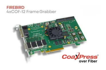
For even faster acquisition and transfer, the recent release of CoaXPress v2.1 means that CXP frame grabbers can support data rates of up to 12.5 Gbps per connection (“CXP-12”), so up to 5 Gbytes/sec in a typical 4-connection system. CoaXPress cables are cost-effective and support data transmission over longer lengths. Our FireBird Quad CXP-12 3PE8 frame grabber supports some of the fastest image acquisition available.
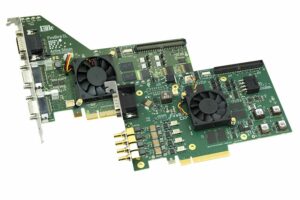
FireBird frame grabbers can also be used in systems to enhance the process of extracting specific features or regions of interest from an image, which can be helpful for diagnosing certain diseases or conditions. In conjunction with GPU processors, our frame grabbers can significantly reduce the time it takes to analyze a large dataset of images. Read more about accelerating processing with a GPU.
Computer vision supports fast and accurate diagnoses
The use of digital slide scanners in pathology has revolutionized the way that pathologists diagnose and study diseases. Digital scanners allow for high-quality images to be captured and shared more easily than with traditional methods, which leads to more accurate diagnoses and better patient outcomes.
Additionally, the use of frame grabbers and embedded vision systems improve image quality, reduce noise, and increase the efficiency of analysis. See how our computer vision components are supporting medical imaging and contact us to discuss your requirements further.